- by Daily Talkin Staff
- July 8, 2026
-

-
-
Loading
Loading
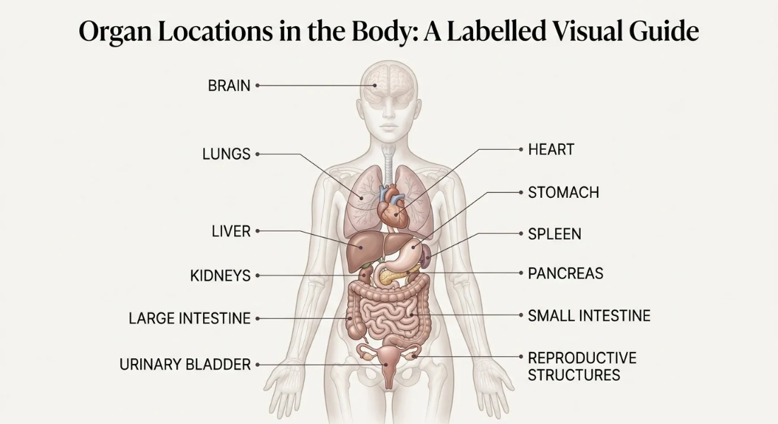
Organ locations in the body can be confusing when labels overlap or the body's left and right sides appear reversed. Knowing where major organs sit makes anatomy diagrams easier to understand and interpret.
This guide uses a labelled diagram, regional explanations and a quick reference table to help you identify the approximate positions of major organs in the head, chest, abdomen and pelvis.
Major organs occupy the head and neck, chest, abdomen and pelvis.
Anatomical left and right refer to the person's own sides, not the viewer's.
The heart sits centrally between the lungs and extends towards the left.
The liver lies mainly in the upper right abdomen; the stomach and spleen lie towards the upper left.
The kidneys sit towards the back of the upper abdomen, one on either side of the spine.
Use front and posterior views together when a diagram obscures an organ's position.

Major organs occupy recognisable positions throughout the head and neck, chest, abdomen and pelvis. Their locations can be described by their relationship to the body's midline, front, back and nearby structures.
A labelled diagram helps you connect these positions with the correct anatomical regions. Labelled front view diagram showing the approximate locations of major human organs.
Front view of major organs in their approximate anatomical positions. A person's left side appears on the viewer's right when looking at the body from the front.
For a broader explanation of human organ types, their functions, and how organs work together across the body explore our complete Human Organs guide → see the full guide here for additional context.
The diagram provides an overview but some organs sit behind others or extend across more than one region. The kidneys for example lie towards the back of the upper abdomen so a front facing illustration may not show their position clearly.
Anatomical location descriptions refer to the body's own left and right sides not the viewer's. Terms such as upper, lower, front and back help describe where an organ sits in relation to other structures. Reading these directions correctly makes a labelled illustration easier to interpret.
Imagine standing face to face with the person in the diagram. Their anatomical left is on your right while their anatomical right is on your left.
This convention remains the same in medical descriptions regardless of which way the illustration faces. Check any orientation arrows before identifying a side specific organ such as the liver or spleen.
These common anatomical terms describe relative positions:
Superior: towards the head or upper part of the body.
Inferior: towards the feet or lower part of the body.
Anterior: towards the front of the body.
Posterior: towards the back of the body.
Medial: closer to the body's midline.
Lateral: farther from the midline towards either side.
For example, the heart is medial to the lungs while the kidneys sit posteriorly in the abdomen. These terms describe relationships rather than exact distances.
The brain sits inside the skull, within the cranial cavity, while the eyes, ears and other major sensory structures occupy identifiable positions around the head.
The neck lies between the head and chest. These landmarks help you orient yourself before moving down a full body diagram.
The brain is inside the skull within the cranial cavity. It occupies most of the space enclosed by the skull rather than sitting in one small area near the forehead or the back of the head.
The forehead marks the front of the head, the temples sit at the sides, and the back of the skull lies behind the brain. These external landmarks help orient the head although they do not show every internal boundary.
The eyes sit towards the front of the head within the eye sockets. The ears are positioned on either side while the nose and mouth occupy the central and lower parts of the face.
The neck begins below the head and continues towards the chest. When reading an illustration, use these visible landmarks to establish orientation before following labels towards deeper structures.
The lungs occupy the right and left sides of the chest within the thoracic cavity. The heart lies between them, near the body's midline with much of it extending towards the left. The rib cage surrounds these structures, and the diaphragm forms the boundary between the chest and abdomen.
The heart lies between the lungs and extends more towards the left side of the chest. The diaphragm separates the chest from the abdominal region.
The heart is not located entirely on the left side of the chest. It sits centrally between the lungs close to the midline with a greater portion extending to the left.
This distinction matters when interpreting simplified diagrams. A heart symbol placed over the left chest may help identify the general region but it does not accurately represent the organ's complete position.
The right lung occupies the right side of the chest, and the left lung occupies the left side. Both sit inside the rib cage with the heart positioned between them.
The lungs extend from the upper chest down towards the diaphragm. Because the heart takes up space towards the left the two sides of the chest are not exact mirror images.
The abdomen lies between the chest and pelvis and contains several major organs at different depths. The liver and stomach occupy the upper abdomen while the intestines fill much of the central and lower region. The kidneys sit farther back on either side of the spine.
The liver lies mainly in the upper right abdomen extending towards the midline. The stomach sits mainly in the upper left abdomen just below the diaphragm.
The gallbladder is tucked beneath the liver. The spleen lies towards the body's left side in the upper abdomen, near the stomach. These positions are approximate, and the organs' shapes mean their boundaries do not align neatly with simple boxes on a diagram.
The pancreas lies deep in the upper abdomen behind the stomach. Its position may be difficult to understand from a front view because other structures can obscure it. The small intestine occupies much of the central and lower abdomen.
The large intestine frames a substantial portion of the abdominal cavity before continuing down into the pelvis. On a labelled illustration follow the outlines and leader lines rather than assuming every structure is fully visible.
The kidneys are a pair of organs towards the back of the upper abdomen one on each side of the spine. They sit behind the main contents of the peritoneal cavity which helps explain why front facing diagrams can make their positions unclear.
Posterior view of the kidneys positioned towards the back of the upper abdomen on either side of the spine. The right kidney usually sits slightly lower than the left because of the position of the liver.
A posterior view is often the clearest way to identify both kidneys and understand their relationship to the spine.
The pelvis is the lower trunk region beneath the abdomen. The urinary bladder sits towards the front, the rectum lies towards the back, and internal reproductive organs occupy positions that vary with individual anatomy.
Their relative locations are easier to understand when the pelvis is viewed separately from the rest of the body.
The urinary bladder sits in the front portion of the pelvis immediately behind the pubic bone. It lies below the main abdominal region and above the pelvic floor.
Its position changes somewhat as the bladder fills so a diagram should be treated as a reference rather than an exact representation of every individual's anatomy.
The rectum lies towards the back of the pelvis. In typical female reproductive anatomy the uterus sits between the bladder and rectum, while the ovaries are positioned on either side of the uterus.
In typical male reproductive anatomy, the prostate sits below the bladder, and the seminal vesicles lie behind it. These are general positional descriptions; individual anatomical relationships can vary.

Some organs are paired across the body's two sides while others sit centrally or mainly towards one side. The heart is near the midline but extends left whereas the liver lies predominantly on the right. This quick reference table summarises the approximate positions of key structures.
Organ or structure | Approximate location |
Right and left lungs | Opposite sides of the chest |
Heart | Central chest, slightly left of the midline |
Liver | Mainly upper right abdomen |
Stomach | Mainly upper left abdomen |
Spleen | Upper left abdomen |
Kidneys | Towards the back of the abdomen, one on each side of the spine |
Small intestine | Central and lower abdomen |
Large intestine | Frames much of the abdominal cavity |
Urinary bladder | Front of the pelvis |
Brain | Within the skull |
The lungs and kidneys are prominent examples of paired internal organs. One lung sits on each side of the chest while one kidney lies on each side of the spine towards the back of the upper abdomen.
Paired organs occupy corresponding sides but their positions are not necessarily identical. Differences in surrounding structures can affect their height, shape and relationship to nearby organs.
The body's internal arrangement is not a perfect mirror image. The liver occupies much of the upper right abdomen, while the spleen lies towards the upper left.
The heart provides another example: it sits near the midline but extends more towards the left. For accurate identification use the organ's full position and nearby landmarks rather than expecting matching structures to sit at exactly the same height.

A two dimensional diagram simplifies three dimensional anatomy. To identify a structure accurately combine its label with the body region and its relationship to nearby landmarks.
Do not rely on a single point on the illustration especially when organs overlap or sit behind other structures.
Organs sit at different depths so a front facing view can make one structure appear to cover another. This is a limitation of viewing three dimensional anatomy on a flat page not necessarily an error in the illustration.
Use a posterior view or another anatomical angle when a structure is obscured. The kidneys are a useful example because their position towards the back of the abdomen is easier to see from behind.
No Anatomy diagrams show typical approximate positions not a precise map that applies identically to every person. Body size, posture and individual anatomical differences can affect the apparent positions and relationships of structures.
Use diagrams to understand general location and orientation. When exact anatomical detail matters for education or clinical work consult a reputable anatomy reference rather than relying on a simplified illustration alone.
Major organs are easiest to locate when you combine the body region, anatomical left and right, and the position of nearby structures. Use the labelled front view for an overview, then check posterior or closer views for organs that sit deeper in the body.
The reference table provides a quick way to confirm approximate positions without confusing a diagram's viewpoint with the body's own orientation.
Major organs occupy the head and neck, chest, abdomen and pelvis. The abdomen contains many of the major organs shown on standard anatomy diagrams.
The stomach and spleen lie in the upper left abdomen. The left lung and kidney occupy the left side while the heart extends left of the midline.
The liver lies mainly in the upper right abdomen. The right lung occupies the right side of the chest, and the right kidney lies beside the spine.
Yes. The heart sits centrally between the lungs, close to the midline although much of it extends towards the left side.
The kidneys lie towards the back of the upper abdomen with one on each side of the spine. A posterior diagram shows them more clearly.
No A labelled diagram shows typical approximate positions and may simplify organ depth, proportions and individual anatomical variation.


Reader Discussion
Leave a Reply