- by Daily Talkin Staff
- July 8, 2026
-

-
-
Loading
Loading
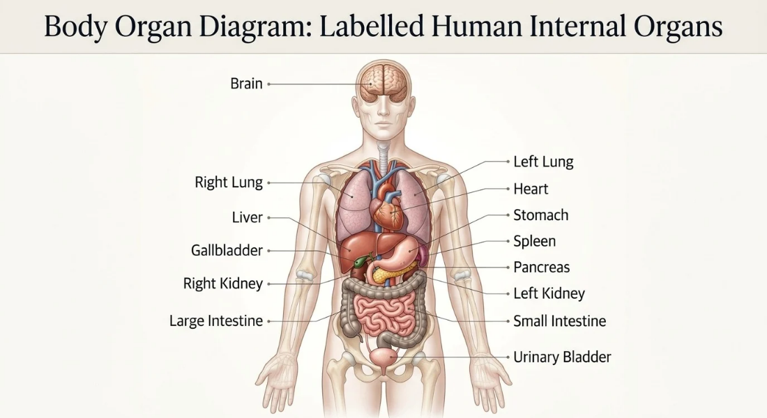
A diagram can make organ names easier to recognise but only when its labels point to the correct structures. A body organ diagram is a labelled visual reference showing major internal organs in their approximate anatomical positions.
This guide explains how to read the illustration, interpret its orientation and check its accuracy and accessibility.
A body organ diagram identifies major internal organs through labels and visual pointers.
Follow each leader line to its endpoint instead of relying on label proximity.
Confirm whether the illustration shows an anterior or posterior view.
Check that similar and overlapping structures have distinct accurate labels.
Use readable typography, descriptive alt text and a caption that matches the actual image.
Treat simplified illustrations as approximate anatomical references not exact representations of every individual.

Place the labelled illustration at the top of this section so readers can identify the structures before reading further. Use a clear front facing human figure with readable callout lines and recognisable organ shapes.
The published image should be original or properly licensed and checked against a reliable anatomy reference. Its labels must match the structures actually shown with no misleading pointers or decorative details that obscure identification.
A full body illustration can label the brain, heart, lungs, liver, stomach, kidneys, pancreas, spleen, small intestine, large intestine and urinary bladder. Include the gallbladder if the artwork depicts it clearly.
The final label list must reflect the actual image. A structure hidden behind another organ should not receive a pointer that appears to identify a different structure.
Use thin high contrast leader lines that end clearly on the intended organ. Keep lines from crossing where possible, and position labels outside crowded areas when necessary.
Consistent typography helps readers distinguish organ names from other annotations. Check the diagram at its intended display size, especially on mobile screens, where small labels and closely spaced pointers can become difficult to follow.
Restrained colours can help distinguish adjacent structures but they should not suggest that an illustration reproduces natural tissue colours exactly. A colour key is useful only when the colours communicate a defined distinction.
Maintain sufficient contrast between organs, labels and the background. Readers must still be able to identify structures through their outlines and labels if they cannot distinguish the colours.
Use a caption such as Labelled human body internal organs diagram showing the approximate positions of major organs in an anterior (front) view. The illustration simplifies three dimensional anatomy for identification.
This wording identifies the visual's purpose, perspective and limitations without repeating every label. Adjust it if the final image uses a different orientation or omits any structures.
To read a labelled illustration correctly, identify its viewing perspective, follow each label or leader line to its endpoint, and match the pointer to the visible structure. Use the caption or legend when a symbol is unclear.
The reference perspective matters because a front facing figure's left and right are defined from the illustrated person's viewpoint not the viewer's. OpenStax explains these conventions in its Anatomical Terminology reference.
Start with the printed organ name then follow its leader line to the endpoint. Check the outline and neighbouring structures to confirm that the pointer reaches the intended organ.
When several labels sit close together do not assume that the nearest text identifies the nearest structure. The pointer's endpoint determines the intended connection. Colour and apparent size can help but neither should replace a clear label.
A front facing illustration shows the body from an anterior view. The artist may remove the front surface in a cutaway or use a transparent representation to reveal internal structures.
The caption should explain which method the image uses. This distinction helps readers understand that the illustration reveals structures normally concealed beneath the body's surface rather than showing them through an ordinary external view.
Read the pointer, label placement and visible outline together. Abdominal structures and intestinal loops can overlap in simplified illustrations making precise callout placement particularly important.
If a small structure cannot be identified clearly at full body scale use a labelled inset. The inset should enlarge the relevant detail without replacing or competing with the main diagram.
Spell out organ names wherever space allows. If abbreviations, numbered markers or arrows are necessary explain them in a compact legend placed near the illustration.
Avoid unexplained colour keys or numbering systems. A reference diagram should support quick identification not require readers to decode several symbols before they can recognise a structure.
Assess a diagram by checking whether its intended structures are recognisable, accurately positioned and labelled individually. Completeness depends on the image's stated scope not an unsupported claim that every diagram must show a fixed number of organs.
For a general full body reference, review the brain, heart, lungs, major abdominal organs, intestines and urinary bladder against the final artwork. Each label should identify a visible structure without implying that simplified illustrations show every anatomical detail.
The brain should appear within the head while the heart and both lungs should be distinguishable in the chest. Use recognisable shapes and ensure the heart's placement reflects its actual anatomical relationship to the lungs.
Avoid drawing the heart as perfectly centred between two identical lungs. Decorative symmetry can make an illustration attractive but less useful as an anatomical reference.
Check the liver, stomach, pancreas, spleen, kidneys and intestines against the artwork. Give the pancreas and spleen clear pointers if larger structures partially obscure them.
Include the gallbladder when it is visible and can be labelled reliably. A simplified diagram may conceal parts of some organs, so do not imply that every structure is equally exposed or easy to see.
The small intestine should be distinguishable from the large intestine's framing course wherever both appear in the image. Their outlines and label endpoints should make the difference clear.
If the illustration conceals part of either structure, adjust the pointer or provide an inset. Avoid a single generic “intestines” label when the diagram claims to identify the two structures separately.
If included the urinary bladder should be depicted in the pelvic region with its label connected directly to the illustrated structure.
Keep its pointer distinct from the kidney labels. If the bladder is absent or too indistinct to identify leave it out of the image's label list rather than suggesting that the diagram shows it accurately.

A labelled illustration converts three dimensional anatomy into a two dimensional reference. Its orientation, proportions and simplified shapes affect how readers interpret the positions of internal structures.
Check the caption before assigning sides or comparing paired organs. The OpenStax anatomy reference explains the standard anatomical position and directional terminology used to avoid this kind of confusion.
In a standard front facing view the illustrated person's right side appears on the viewer's left. The person's left therefore appears on the viewer's right.
State this orientation in the caption or a nearby note. Without that clarification readers may assign sides according to their own perspective instead of the body's anatomical orientation.
The lungs and kidneys are examples of paired organs that may appear in a full body illustration. Show each member clearly enough for readers to distinguish it from the other.
Paired organs often have similar forms but they are not necessarily identical or positioned as perfect mirror images. The diagram should communicate their approximate placement without forcing artificial symmetry.
Educational illustrations may simplify proportions, separate overlapping structures or omit details to make labels easier to read. They represent anatomy selectively rather than reproducing every feature of an individual body.
Organs can also vary in shape and exact position between individuals. Treat the drawing as a reference for approximate placement not an exact map of every person's internal anatomy.
A posterior, or back, view can help when a structure is difficult to interpret from the front. Add it as a small clearly labelled inset only when it provides useful information beyond the main illustration.
View | Main purpose |
Anterior (front) | Provides the primary full body reference for visible internal structures. |
Posterior (back) | Offers an additional perspective when it helps clarify a structure's position or appearance. |
Keep the front view dominant unless the second perspective genuinely improves identification.

A reliable visual reference needs accurate organ placement, readable labels and an accessible text alternative. Its appearance alone does not establish anatomical accuracy verify the illustration against a credible source and ensure its annotations identify the structures correctly.
Compare the image's labels, orientation and organ positions with a suitable anatomy reference such as OpenStax Anatomy and Physiology 2e or the MedlinePlus Medical Encyclopedia.
Where feasible, ask a qualified anatomy educator or appropriate clinical reviewer to assess the final artwork. Publish reviewer credentials only when a genuine review has taken place.
Use readable text, strong contrast, uncluttered leader lines and sufficient spacing between labels. Test the image at mobile size as well as desktop size since small embedded text can become illegible on narrow screens.
Provide descriptive alt text and a nearby text equivalent identifying the depicted organs. Essential information should not depend solely on colour, tiny lettering or visual interpretation.
Alt text should describe the actual illustration, its viewing angle and the principal organs visibly labelled. Include “body organ diagram” naturally when it accurately describes the image.
The caption can explain the simplified representation and approximate positions in greater detail. Do not list organs that the image omits, and avoid repeating every label in both the alt text and caption.
A simplified diagram supports general learning and visual identification but it cannot determine an individual's internal anatomy or diagnose a medical condition.
Use a detailed educational reference when you need more anatomical information. This is part of our complete Human Organs guide → see the full guide here.
A labelled body organ diagram is most useful when its structures are recognisable its pointers reach the correct organs and its viewing orientation is clear. Simplified illustrations show approximate positions rather than every anatomical variation.
Use the visual as an educational reference supported by an accessible text alternative, and check its labels before relying on it for identification.
A body organ diagram is a labelled visual reference showing major internal organs in their approximate positions. It helps readers identify structures without requiring lengthy anatomical explanations.
A general illustration may show the brain, heart, lungs, liver, stomach, kidneys, pancreas, spleen, intestines and urinary bladder. The actual labels depend on the structures depicted.
Identify the viewing perspective, follow each label or leader line to its endpoint, and match the pointer to the visible structure. Check the legend if a symbol is unclear.
Not usually in a standard front view. The illustrated person's right appears on your left while their left appears on your right. Check the orientation note to confirm.
No Organ shapes and exact positions vary between individuals. Educational illustrations simplify anatomy to make structures easier to identify and should be treated as approximate references.
No A general educational diagram cannot establish an individual's internal anatomy or diagnose a condition. Use appropriate medical assessment when you have a health concern.


Reader Discussion
Leave a Reply